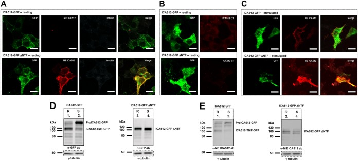
FIG 6.
NTF targets ICA512 to SGs. (A to C) Confocal microscopy images of resting (A and B) or HGHK-stimulated (C) INS-1 cells transfected with ICA512-GFP (top panels; green) or ICA512-GFP ΔNTF (bottom panels; green). Live cells were incubated at 4°C with the mouse anti-ME ICA512 antibody (A and C), together with the guinea pig anti-insulin antibody in panel A, or were incubated with the mouse anti-ICA512 CT antibody (B). After fixation, the binding of the primary antibodies to the cell surface was detected by incubation with Alexa Fluor-conjugated anti-mouse (A to C) (red) or anti-guinea pig (A) (white) IgG. Merged images are additionally shown for panels A and C. Bars = 10 μm (n ≥ 3). (D and E) Immunoblotting for GFP (D) and ME ICA512 (E) in lysates of resting (R) or HGHK-stimulated (S) INS-1 cells transfected with ICA512-GFP or ICA512-GFP ΔNTF (n ≥ 3). For normalization, the same lysates were also immunoblotted for γ-tubulin.

