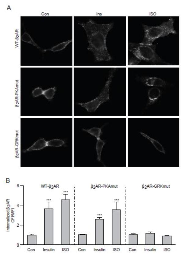
Figure 2. Insulin and ISO induce internalization of β2AR in H9c2 cardiac myoblasts.
H9c2 myoblasts expressing wild type (WT), mutant β2AR lacking PKA phosphorylation sites (PKAmut), or mutant β2AR lacking GRK phosphorylation sites (GRKmut) were stimulated with insulin (100 nM, 10 min) or ISO (100 nM, 5 min). (A) The distribution of β2AR was examined with immunofluorescence staining. (B) The internalization of β2AR was quantified with Fiji image-processing package. n = 10; *** p < 0.001 when compared to control by one-way ANOVA followed by Tukey’s test.

