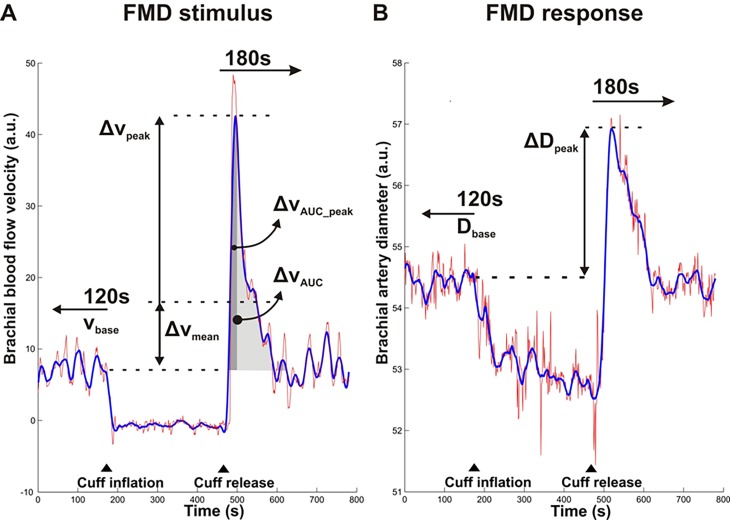
Figure 1. Definitions of baseline and hyperemic brachial artery blood flow velocity and diameter parameters for the assessment of flow-mediated dilation.
Blood flow velocity (A) and diameter curves (B) obtained by beat-to-beat analysis of Duplex video images. Raw data curves (red) were smoothed (blue) to reduce noise, particularly in the peak change estimates. Baseline blood flow velocity (vbase) and diameter (Dbase) were determined over the 120 seconds prior to cuff-inflation. The peak change in blood flow velocity (Δvpeak) was determined as the maximum velocity reached within 180 seconds after cuff-release while the mean change in blood flow velocity (Δvmean) was determined as the average velocity over the 180 seconds after cuff-release, both minus the baseline blood flow velocity vbase (A). The peak change in diameter (ΔDpeak) was determined as the maximum diameter reached within 180 seconds after cuff-release minus the baseline diameter Dbase (B). Indicated also are the full area under the curve of the hyperemic velocity curve above baseline level (ΔvAUC) and the area integrated till peak-time of velocity curve (ΔvAUC_peak). ▲indicates the timing of rapid cuff inflation and release.

