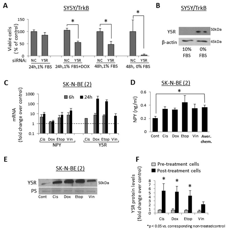
Figure 5. NPY system is up-regulated in NB cells under pro-apoptotic conditions.

A. SY5Y/TrkB cells were transfected with negative control (NC) or Y5R siRNAs and cultured in 1% or 0% FBS culture media for 24h–48h, with or without doxorubicin (1μg/ml). Cell viability was measured by MTS assay. B. SY5Y/TrkB cells were cultured for 24h in 10% or 0% FBS media and protein levels of Y5R were detected by Western blot. C. SK-N-BE(2) cells were cultured for 24h in 1% FBS media and then treated with four different chemotherapeutics – cisplatin (Cis, 0.5μg/ml), doxorubicin (Dox, 0.5μg/ml), etoposide (Etop, 2.5μg/ml) or vinblastine (Vin, 1μg/ml) for 6 or 24h. mRNA levels of NPY and Y5R were measured by real-time RT-PCR. D. SK-N-BE(2) cells were treated with chemotherapy as above and NPY released to the cell culture media was measured by ELISA. E. Protein levels of Y5R were assessed by Western blot in chemotherapy-treated SK-N-BE(2) cells. F. A panel of NB cells derived from patients before (CHLA-15, SMS-KCN, SMS-KAN) and after chemotherapy (SK-N-BE(2), CHLA-20, SMS-KCNR, SMS-KANR) was treated for 6–12h with chemotherapeutic agents, as above, and Y5R detected by Western blot. Expression levels from at least three independent experiments for each cell line were quantified by densitometry and the results for all pre- and post-treatment cells were averaged. PS – unspecific protein staining.
