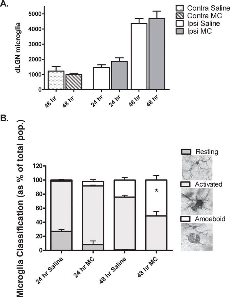
Fig. 1. Minocycline does not inhibit accumulation of microglia but is associated with an increased frequency of amoeboid microglia in the ipsilateral dLGN 48 hours after cortical ablation in P10, MT−/− mice.

A. Using unbiased stereological methods, the number of Iba1 positive microglia in the contralateral (contra) and ipsilateral (ipsi) dLGN were estimated by an investigator blinded to the treatment condition. No significant difference was found between numbers of microglia in saline (Sal) vs. minocycline (MC) treated mice at any time point after injury. *P=0.03, saline vs. minocycline, Mann-Whitney U, N= 3 /contra group, N= 5 /ipsi group. B. An investigator blinded to treatment condition classified Iba1 positive microglia as resting, activated or amoeboid according to morphological criteria (Dailey and Waite, 1999; Ito, et al., 2001; Kreutzberg, 1996). Resting microglia were recognized by a small cell body and numerous, highly ramified processes. Activated microglia were distinguished by an enlarged cell body and short, stout processes. Spheroid microglia with few or no processes, were classified as amoeboid. Stacked columns show each morphology as a percentage of total population with time after injury and treatment. No significant difference was determined between treatment groups at 24 hrs. Percentage of amoeboid microglia was increased in minocycline (MC) vs. saline (Sal) treated mice at 48 hrs, *P= 0.03 Mann-Whitney U, N = 4/ group.
