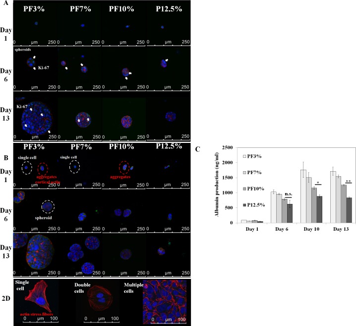
Fig 5. Time dependent spheroid formation and albumin expression inside Huh7.5 cell-laden hydrogels at day 1, day 6, and day 13.
Spheroid growth was observed in Huh7.5 cell-laden hydrogels through F-actin, nucleus proliferation marker (Ki-67), and albumin staining. A. blue: nucleus; red: F-actin; green: Ki-67. B. blue: nucleus; red: F-actin; green: albumin. After spheroids were formed, cortical type red F-actin could be observed among Huh7.5 cells. On the contrary, Huh7.5 cells cultured on 2D glass slide displayed well developed actin stress fibers in single and double cells. C. ELISA for quantification of albumin secretion from each hydrogel with Huh7.5 cells (1x10⁵ cells) initially loaded was performed. One way ANOVA, n = 3: P < 0.01, P < 0.05 and P < 0.001 at day 6, day 10, and day 13, respectively among PF3%, PF7%, and PF10%; t-test (n = 3 between PF10% and P12.5%): *: P < 0.05 at day 10; **: P < 0.001 at day 13; n.s.: not significant.

