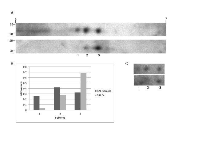
Fig 7. 2D Western blot for TXNPx in amastigotes from BALB/c and BALB/c nude mice.
A. Western blot image showing three isoforms (1, 2, 3) in soluble extracts from BALB/c nude (upper image) and BALB/c (lower image) derived amastigotes. B. Relative abundance of each isoform after normalization (considering the sum of three isoforms similar in the two samples). C. Western blot image showing labeling of the isoforms with anti-phospho S, T, Y pooled antibodies in the membranes shown in A.

