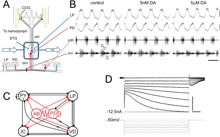
Fig 1. The experimental model.
(A) In situ preparation. The STNS is dissected & pinned in a dish. The commissural ganglia (CoGs) contain DA neurons that project to the STG (black) and L-cells, which are the source of neurohormonal DA (gold). The well surrounding the STG (blue rectangle) is continuously superfused with saline (in/out arrows). There are ~30 neurons in the STG; 2 are drawn: pyloric dilator (PD), lateral pyloric (LP). Network neurons interact locally within the STG neuropil and can project axons to striated muscles surrounding the foregut. The diagram shows that PD & LP neurons project their axons through identified nerves to innervate muscles (rectangles). (B) Spontaneous pyloric network output. The top 2 traces are intra-cellular recordings from the in situ preparation diagrammed in A. The bottom 2 traces represent extra-cellular recordings from identified motor nerves containing pyloric neuron axons. The spikes from three pyloric neurons are indicated on lvn. These simultaneous recordings demonstrate the spontaneous, recurrent, rhythmic motor pattern produced by the circuit; scale bars, 10mV & 500ms. (C) The pyloric circuit. The pyloric network comprises 14 neurons. The diagram represents pyloric neuron interactions within the STG. Open circles represent the 6 cell types, numbers indicate more than 1 cell within a cell type: anterior burster (AB), inferior cardiac (IC), ventricular dilator (VD); pyloric constrictor (PY); filled circles, inhibitory chemical synapses; resistors & diodes, electrical coupling; red, pacemaker kernel and its output connections. (D) Two electrode voltage clamp experiment. Top: Typical LP Ih recording; Bottom: voltage protocol; scale bars, 500ms and 5nA.

