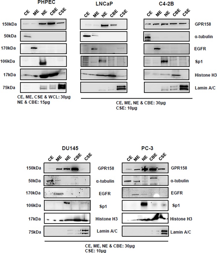
Fig 6. GPR158 subcellular localization in PCa cell lines and PHPECs.
Subcellular protein fractionation was carried out in LNCaP, C4-2B, PC-3 and DU145 cells, as well as PHPECs. The amount of protein loaded for each fraction is indicated on the panels. Western blotting was performed using anti-ICD GPR158 antibody. Specific protein markers were used to validate and confirm the purity of the five subcellular fractions examined: cytoplasmic extract (CE) = alpha-tubulin, membrane extract (ME) = EGFR, soluble nuclear extract (NE) = Sp1, chromatin-bound nuclear extract (CBE) = histone H3 and insoluble cytoskeletal extract (CSE) lamin A/C. The percentage of GPR158 protein present in each fraction was calculated with respect to the total amount of protein in all fractions using Image J analysis of protein band intensities. The data are representative of two independent experiments.

