
Figure 1.
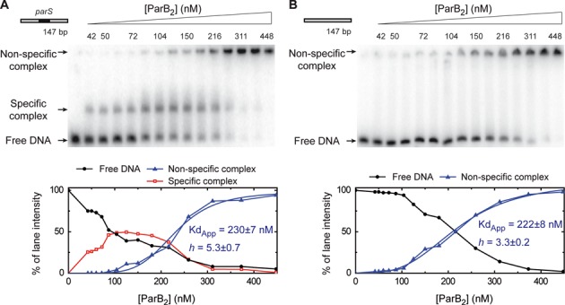
Specific binding of ParB to the parS sequence. Electrophoretic mobility shift assay of ParB binding to a radiolabelled 147-bp substrate in a magnesium acetate containing gel-running buffer. (A) Titration of ParB on DNA containing a single parS site in the centre. (B) ParB titration on an equivalent substrate that is lacking a parS site (see Supplementary Table S1 for details). The species assigned as specific and non-specific complexes are labelled. The lower panels show the quantification of the gels revealing a highly sigmoidal pattern for non-specific binding. These data were fit to Equation (1) to yield the values shown.