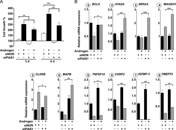
Figure 2.

PIAS1 influences proliferation of VCaP cells. (A) VCaP cells were transfected with siNON (control) or siPIAS1 and cells were exposed to androgen or vehicle and cell numbers were measured by CellTiter96 Aqueous cell proliferation assay as described in ‘Materials and Methods’ section. Growth percentages represent the means of three independent experiments ± SDs. (B) Effect of PIAS1 depletion on the expression of select AR target genes linked to cell growth and apoptosis. Cells were treated as in Figure 1, but RNA quantification was carried out by RT-qPCR with specific primers for BCL6, ATAD2, NR5A2, MAGEH1, CLDN8, MAFB, TNFSF10, CSRP2, IGFBP-3 and TMEFF2 mRNAs. Measurements were normalized to GAPDH mRNA levels, and fold changes were calculated in reference to siNON and vehicle samples. Data points indicate the means of at least three biological replicates ± SDs. Student's t test was used to determine the significance of fold change differences between siNON and siPIAS1 cells when comparing androgen- and vehicle-exposure (***P < 0.001, **P < 0.01 and *P < 0.05).
