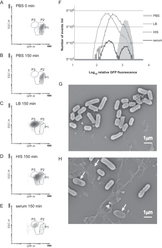
FIG 3.
A bacterial population that survives serum treatment is enriched in rapidly dividing and nondividing cells. Stationary-phase CFT073 cells [carrying plasmid pETgfp-mut2AGGAGG(3)] were diluted in PBS, LB medium, 50% HIS, or 50% serum. The cells were incubated at 37°C without shaking for 150 min, and their fluorescence was measured by using flow cytometry. Density blots of GFP fluorescence (GFP-H) and the SSC parameter (SSC-H) are shown for cells in PBS at zero time (A), in PBS after 150 min (B), in LB medium (C), in HIS (D), or in serum (E). Each dot represents the fluorescence of a single event (particle). (F) The distribution of the fluorescence level of events (single cells) analyzed by flow cytometry is presented as a histogram consisting of 376 repartition bins. The number of events with the respective GFP fluorescence levels in 1-ml cell cultures grown under particular conditions is shown. (G and H) Scanning electron micrographs illustrate the morphology of the E. coli cells after 150 min of incubation in 50% HIS (G) and in 50% serum (H). Arrows indicate the different bacterial cells during lysis.

