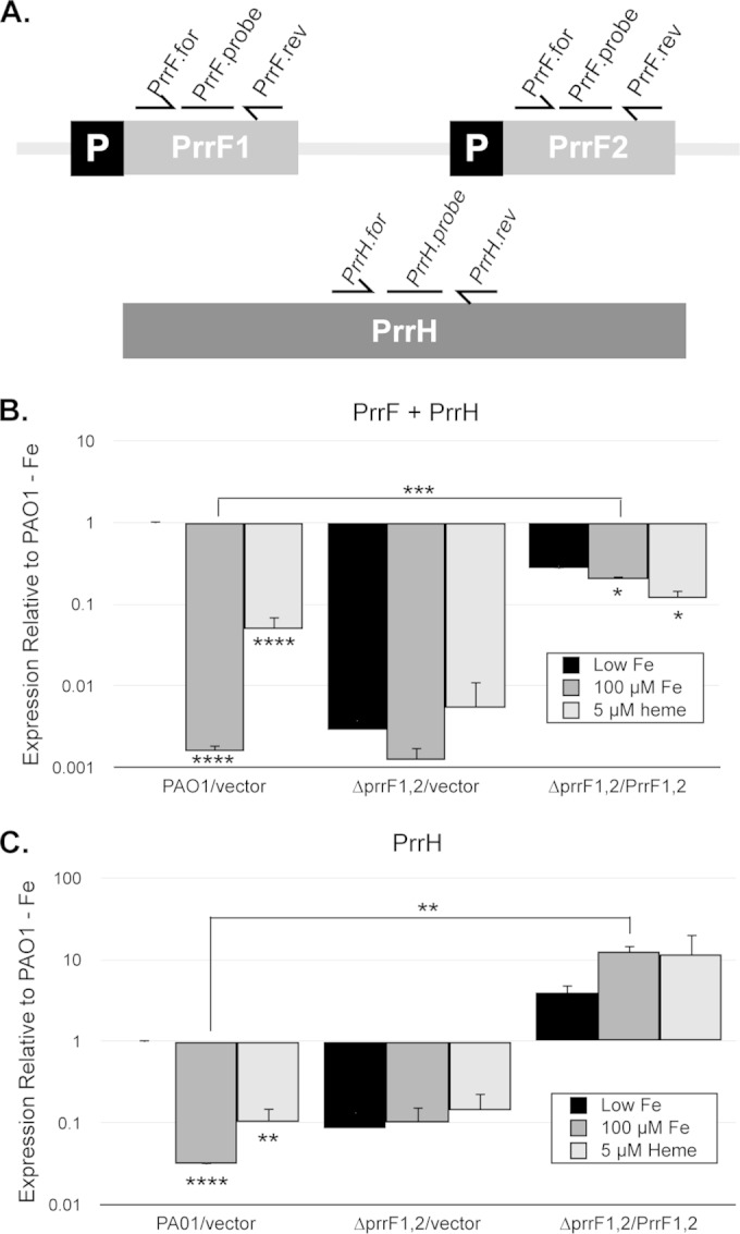
FIG 2.

Iron and heme regulation of the PrrF and PrrH sRNAs in M9 media. (A) Map of the prrF locus, showing approximate locations of the PrrF.for and PrrF.rev primers and PrrF TaqMan probe, which detect the PrrF and PrrH sRNAs (B), and the PrrH.for and PrrH.rev primers and PrrH TaqMan Probe, which only detect PrrH (C). (B and C) The indicated strains were grown for 4 h in M9 medium with 60 μg/ml of tetracycline to deplete intracellular iron stores and then subcultured into fresh M9 medium without iron supplementation (black bars), with 100 μM FeCl3 supplementation (dark gray bars), or with 5 μM heme supplementation (light gray bars). Relative expression of PrrF and PrrH combined (B) and of PrrH (C), detected using the primers shown in panel A, was determined by standard curves as described in Materials and Methods, and expression of each sample was normalized to the PAO1/vector low-iron sample. Error bars indicate the standard deviations from three independent experiments. Asterisks indicate the following P values as determined by two-tailed Student's t test when comparing values upon heme or iron supplementation to that of the low-iron sample, or as otherwise indicated: *, P < 0.05; **, P < 0.001; ***, P < 0.0001; ****, P < 0.00001.
