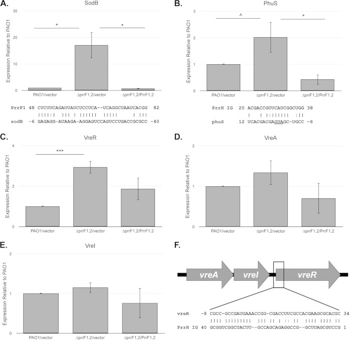
FIG 3.
The ΔprrF1,2 mutant shows altered expression of genes involved in heme homeostasis and virulence. (A to E) The indicated strains were grown for 4 h in M9 medium with 60 μg/ml of tetracycline to deplete intracellular iron stores and then subcultured into fresh M9 medium without iron supplementation. Relative expression of sodB (A), phuS (B), vreR (C), vreA (D), and vreI (E) was determined by standard curves as described in Materials and Methods, and expression of each sample was normalized to the PAO1/vector sample. Primers and probes used are shown in the supplemental material. Error bars indicate the standard deviations from three independent experiments. Complementarity between PrrF1 and the sodB mRNA as previously reported (32) and the PrrH unique region (“PrrH IG”) and the phuS mRNA are shown below the graphs in panels A and B, respectively. The translational start site of phuS is underlined. Asterisks indicate the following P value as determined by two-tailed Student's t test when comparing values upon heme or iron supplementation to that of the low-iron sample, or as otherwise indicated: ∧, P < 0.1; *, P < 0.05; **, P < 0.001; ***, P < 0.0001. (F) Map of the vreAIR operon, indicating the approximate location of complementarity with the PrrH unique region (PrrH IG).

