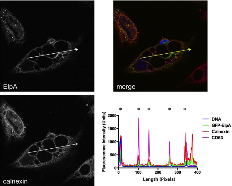
FIG 6.
The PV is decorated with ElpA and calnexin during C. burnetii intracellular growth. Full-length GFP-ElpA (green) was ectopically expressed in HeLa cells that were infected for 30 h with the Dugway isolate. At 18 h posttransfection, cells were processed for fluorescence microscopy. The ER was detected using antibody against calnexin (red) and DAPI-stained DNA (blue). *, PV membrane. Fluorescence intensity analysis (arrows) shows that GFP-ElpA and calnexin are enriched around the PV membrane, as indicated by overlap with the PV marker CD63.

