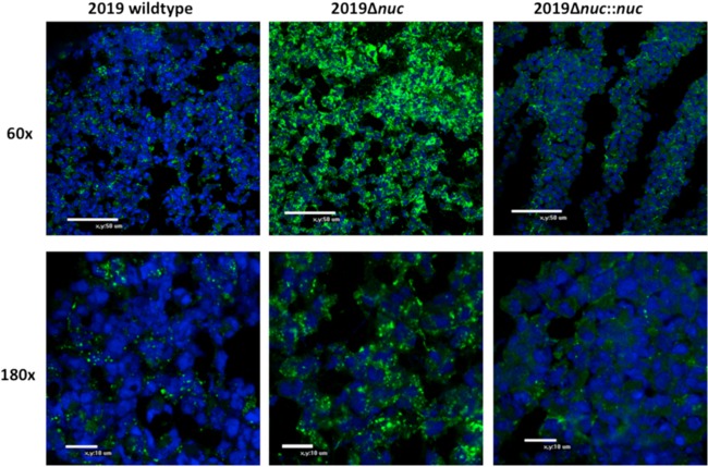
FIG 7.
Confocal microscopy analysis of 10-μm-thick sections of biofilm formation at day 5 in the chinchilla middle ear after infection with NTHI 2019, NTHI 2019Δnuc, and NTHI 2019Δnuc::nuc. The NTHI are stained with MAb 6E4 (green), and DNA is stained with DRAQ5 (blue). There is an aggregation of organisms staining with MAb 6E4 in NTHI 2019Δnuc (B) compared to the more diffuse display of organisms in the wild type and the complemented mutant (A and C).

