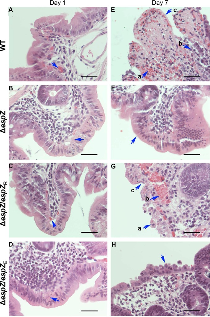
FIG 5.

Infection with the ΔespZ REPEC strain causes minimal and unsustained gut tissue damage. (A to D) H&E-stained sections of rabbit cecum on day 1 postinfection. Shown are representative images from a minimum of three animals per group infected with WT REPEC (A), the ΔespZ mutant (B), or the ΔespZ mutant cis-complemented with REPEC espZ (espZ/espZR) (C) or EPEC espZ (espZ/espZE) (D). Arrows are positioned over sites of proprial or submucosal hemorrhage (a to c) or over apoptosing enterocytes (d). Bar, 50 μm. (E to H) H&E-stained sections of rabbit cecum on day 7 postinfection. Shown are representative images from a minimum of three animals per group infected with either WT REPEC (E), the ΔespZ mutant (F), or the ΔespZ mutant cis-complemented with REPEC espZ (espZ/espZR) (G) or EPEC espZ (espZ/espZE) (H). Arrows are positioned over sites of hemorrhage (Ea, Ec, and Gb), breached or damaged epithelial cells (Eb, Ga, and H), epithelial cells coated with bacteria (Gc), or epithelial cells with normal morphology (F). Bar, 50 μm. Tissue alterations were scored by an American College of Veterinary Pathologists board-certified pathologist (M. W. Riggs) blind to treatment conditions.
