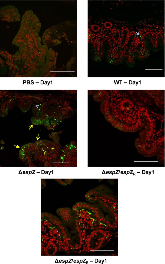
FIG 7.
Infection with the REPEC ΔespZ strain results in significant intestinal cell apoptosis in vivo. Shown are images from TUNEL staining of cecal tissues from rabbits mock gavaged with PBS or infected with the parent WT strain, the ΔespZ mutant, or the ΔespZ mutant cis-complemented with REPEC espZ (espZ/espZR) or EPEC espZ (espZ/espZE). Images are representative of those from multiple fields and multiple animals and highlight the intestinal cell apoptotic status on day 1 postinfection (yellow arrows), when histopathological observations revealed alterations for all groups (Fig. 5). Host cell nuclei were stained red by using the fluorescent marker DAPI, and TUNEL-positive apoptotic cells were stained green by using resorcinol phthalein (fluorescein). Light blue arrows pointing to diffuse staining are subepithelial autofluorescing erythrocytes. Bar, 100 μm.

