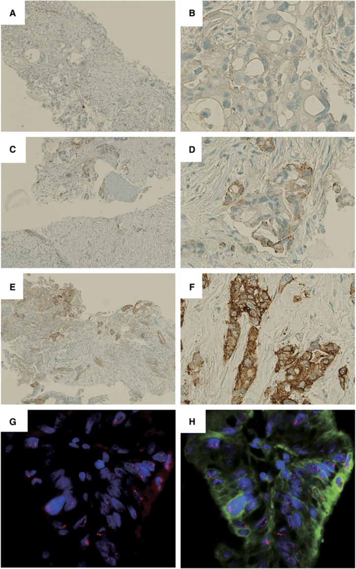
Figure 1.
Immunohistochemical (IHC) and fluorescence in situ hybridisation (FISH) analyses of representative actinin-4 protein expression and ACTN4 copy number, respectively, in LAPC biopsy specimens. (A–F) Immunohistochemical analysis of actinin-4 protein expression. Representative cases of no expression (A, B), weak expression (C, D) and strong expression (E, F) of actinin-4 protein in LAPC cells. (A), (C) and (E) are low-magnitude images. (B), (D) and (F) are high-magnitude images of regions of (A), (C) and (E), respectively. (G, H) Fluorescence in situ hybridisation analysis of representative cases with a copy number increase (CNI) in ACTN4.

