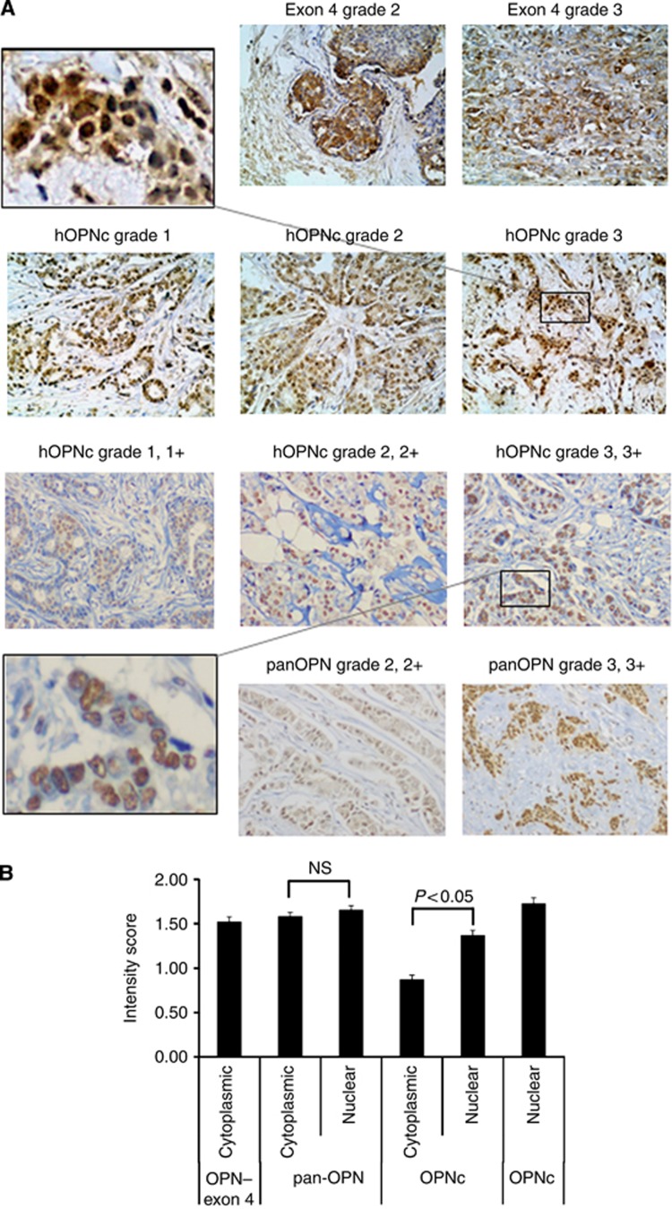
Figure 1.
Osteopontin staining. (A) (top row) Cytoplasmic staining for Osteopontin–exon 4 in invasive ductal carcinomas (histopathological grades 2–3, and staining intensity 2–3) from the Polish cohort. (Second row) Nuclear staining of Osteopontin-c in invasive ductal carcinomas (histopathological grades 1–3, staining intensity 1–3) from the Polish cohort. The insert in the top row (left) shows a zoomed-in picture of the grade 3 staining. (Third row) Anti-Osteopontin-C staining of invasive ductal carcinomas (histopathological grades 1–3, staining intensity 1–3) from the Swedish cohort. The insert in the bottom row (left) shows a zoomed-in picture of the grade 3 staining. (bottom row) Anti-pan-Osteopontin staining in invasive ductal carcinomas from the Swedish cohort. For all pictures, counterstaining with hematoxilin was performed and the original magnification was × 200. (B) Mean values and s.e. of the immunohistochemistry intensity scores for pan-Osteopontin, Osteopontin-c and Osteopontin–exon 4 are shown (for clarity, per cent positivity is not shown). The results for the Swedish cohort and Polish cohort (far left and right bars in the graph) are shown separately. Osteopontin–exon 4 is exclusively cytoplasmic, Osteopontin-c is predominantly nuclear and pan-Osteopontin distributes in both compartments. The differences between nuclear and cytoplasmic staining intensity were assessed by t-test and a P-value<0.05 was considered significant. NS, not significantly different.

