
Figure 3.
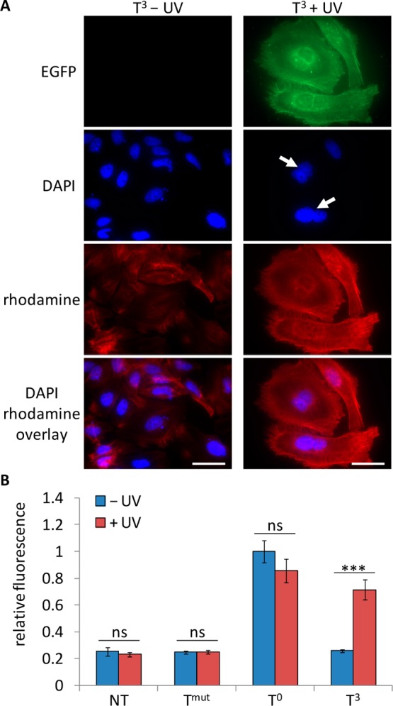
(A) Light-induced expression of Plk3. HeLa cells were transfected with the T3-caged EGFP-Plk3 plasmid followed by irradiation of the caged construct (365 nm, 5 min, 25 W) and incubated for 48 h. The cells were then fixed and stained with DAPI (nuclei) and rhodamine phalloidin (actin filaments) prior to imaging (63× magnification). White arrows indicate binucleated cells, and scale bars indicate 50 μm. (B) Light-induced activation of caspase-3. HeLa cells were transfected with the Tmut negative control, T0-noncaged, and T3-caged EGFP-Plk3 plasmids. The cells were either irradiated (365 nm, 5 min, 25 W) or kept in the dark and lysed after 48 h. The lysate was assayed with a fluorogenic caspase-3 substrate (Calbiochem). Fluorescence units were normalized to the noncaged control, and standard deviations were calculated from three individual experiments. ns = not significant (P > 0.05), *** = highly significant (P < 0.001).