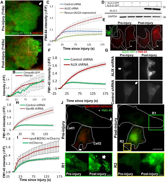
Figure 6. Ca2+-triggered ESCRT/Vps4B assembly allows cell membrane repair.
Following laser injury of myoblasts in presence of FM dye, repair of cell membrane was monitored by following dye entry, which results in increase in FM-dye fluorescence. Cellular dye fluorescence was normalized to the time point prior to injury (ΔF/F). (A) Live cell TIRF (green) and widefield (red) images of a myoblast expressing Chmp4B-GFP (green) and injured in the presence of FM4-64 dye (red) by laser (arrow). (B) The plot shows kinetics of cell membrane repair (FM 4-64 intensity) and Chm4B-GFP accumulation at the injured cell membrane. (C) Plot showing FM 4-64 entry following laser injury of 10-12 cells transfected for 72 hours with control siRNA (blue line) or ALG-2 siRNA with no subsequent treatment (red line) subsequent transfection with siRNA resistant ALG2-GFP plasmid (ALG2r-GFP). (D) Western blot analysis of expression of ALG2-GFP and ALG2r-GFP in cell lysates from HeLa cells transfected with these plasmids with or without ALG-2 siRNA and probed with ALG2 antibody. Bands corresponding to the GFP-tagged protein are shown here and the uncropped blots are shown in Supplementary Fig. 5C (E) Images showing laser injury-induced uptake of FM4-64 dye (red) in HeLa cells expressing or not expressing ALG2r-GFP (inset, green cell). (F) Kinetics of injury-triggered FM 1-43 entry in stable clones of HeLa cells expressing ALIX shRNA (red symbol) or empty vector (control; green symbol) averaged for 10-15 cells in each condition. (G) Representative images of a pair of ALIX shRNA or empty vector control cells injured (at the site marked by the arrow) in presence of FM 1-43 dye. (H) Kinetics of injury-triggered FM 1-43 entry in stable clones of HeLa cells expressing Vps4B shRNA (red symbol) or empty vector (control; green symbol) averaged for 10-15 cells in each condition. (I) Kinetics of injury-triggered FM 1-43 entry in up to 15 C2C12 myoblasts each transiently expressing mCherry (green) or Vps4B(DN)-mCherry. (J) Representative images of FM1-43 dye entry following injury of a pair of C2C12 myoblasts expressing (Cell2) or not expressing (Cell1) Vps4B(DN)-mCherry for 24 hours. Zoomed inset R1 and R2 show the region near the site of injury for cell 1 (lacking) and cell 2 (expressing) Vps4B(DN)-mCherry. Arrow in R1 shows the site where the damaged membrane was excised from the repaired cell and the diffraction limited dots represent the vesicles released during excision of the membrane. Error bars represent SE, and scale bars represent 10μm for the whole cell images and 2μm for the zoomed regions R1 and R2.

