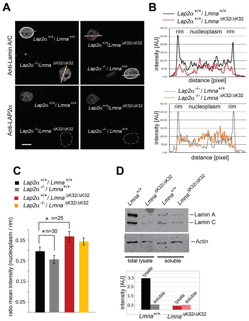
Fig. 2. Lamin A/C protein is redistributed to the nuclear interior in LmnaΔK32/ΔK32 fibroblasts in a LAP2α-independent manner.
(A) Co-cultures of primary dermal fibroblasts with indicated genotypes isolated from new-born littermates were processed for confocal immunofluorescence microscopy. Cells were stained with antibodies to lamin A/C and LAP2α, with the latter allowing identification of the genotype in mixed cultures. Scale bar denotes 10 μm. (B) Quantitation of intranuclear lamin staining was done by fluorescence intensity measurements along the dashed line shown in image using the profile tool in Zeiss LSM Image Browser. (C) Ratios of nucleoplasmic over peripheral mean A-type lamin fluorescence intensity were plotted in the histogram. In LmnaΔK32/ΔK32 fibroblasts, the ratios are significantly increased versus wild-type controls (n=25, P-value<0.05). In the Lmna+/+ background, nucleoplasmic lamins are lost and the ratio decreases significantly upon loss of LAP2α (n=30, P-value<0.05). (D) Immunoblots of total cell lysates and of soluble cell fractions following lysis of cells in Hepes buffer plus 0.5% Triton X-100 and 0.1% SDS, probed for lamin A/C and actin. Protein bands were quantified by ImageJ.

