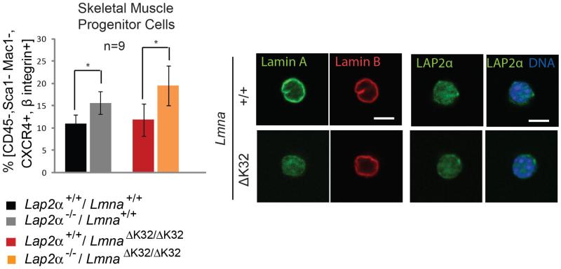
Fig. 5. Lap2α−/− -specific increase in SMPCs is not affected in LmnaΔK32/ΔK32 mice.
Skeletal muscle progenitor cells (SMPCs) (CD45-/Sca1-/Mac1-/CXCR4+/β1-integrin+) were obtained from skeletal muscles (Gastrocnemius, Soleus, Tibialis anterior, Quadriceps, Triceps) and analyzed by flow cytometry or processed for confocal immunofluorescence microscopy. The number of CD45-Sca1-Mac1-CXCR4+β1-integrin+ (SMPC) cells within the parent population (CD45-Sca1-Mac1-) is presented in left panel. Lap2α−/− mice show a significant increase in SMPCs in comparison to their wild-type littermates (n=9, P-value=0.03 as determined by Student’s t-test). Similarly double mutant LmnaΔK32/ΔK32 / Lap2α−/− mice show an increase in SMPCs in comparison to their single mutant LmnaΔK32/ΔK32 littermates (n=9, P-value=0.02 as determined by Student’s t-test). (Right panel shows immunofluorescence microscopic confocal images of isolated SMPCs stained for lamin A/C, lamin B and LAP2α. Scale bar denotes 5 μm.

