
Figure 2.
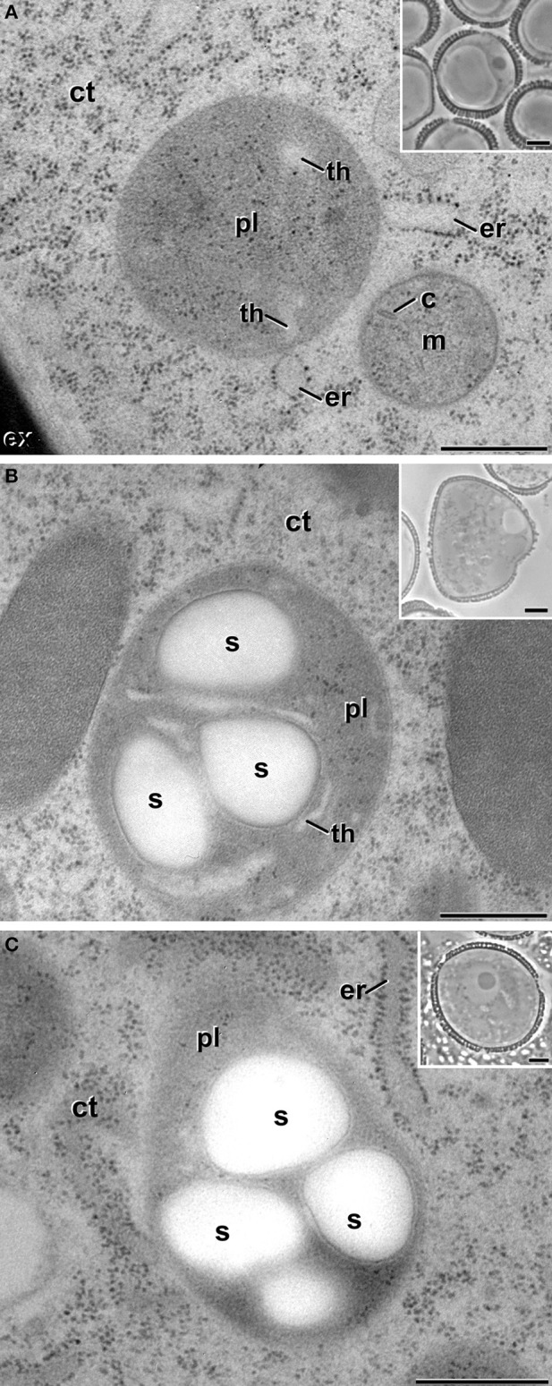
Plastids (pl) of non-embryogenic B. napus cells. (A) Proplastid of a vacuolate microspore during in vivo development within the anther. (B) Amyloplast of an in vitro cultured, pollen-like structure. (C) Amyloplast of a pollen grain within the anther. Insets show light microscopy sections of the corresponding stages. (C), mitochondrial crista; ct, cytoplasm; er, endoplasmic reticulum; ex, exine; m, mitochondria; s, starch; th, thylakoid. Bars: 500 nm, insets: 5 μm.