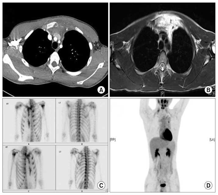
Fig. 1.
(A) A chest computed tomography (CT) scan illustrating a mass in the anterior chest wall without mineralization. (B) A magnetic resonance image displaying an extensive highly vascular extraosseous mass. (C) A whole-body bone scintigraphy image. (D) A positron emission tomography-CT scan showing a diffuse infiltrative lesion with bony destruction and mild uptake.

