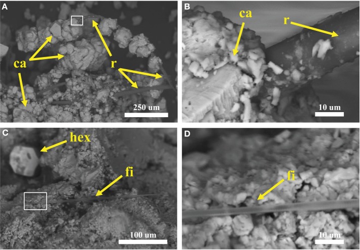
Figure 6.
Scanning electron microscopy (SEM) of samples collected at Manleluag Spring National Park. (A) Terrace samples consist of rhomboid calcite crystals (ca), shown here precipitating on organic matter, likely a plant root (r). (B) Close-up of box in (A). (C) Putative cyanobacterial filament (fi) interbedded with calcite crystals (note hexagonal crystal, hex, in background). (D) Close-up of box in (C).

