FIGURE 1.

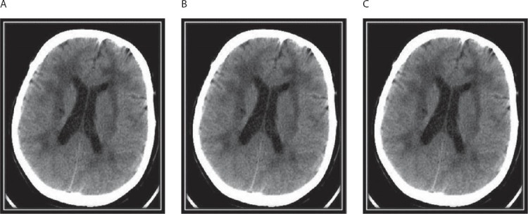
CT scan showing multiple paraventricular and deep white matter hypodensities (A,B), lacunar infarcts in the left nucleus lentiformis (B) and the pons (C), hypodensities in the capsula externa and anterior temporal lobe (C).

CT scan showing multiple paraventricular and deep white matter hypodensities (A,B), lacunar infarcts in the left nucleus lentiformis (B) and the pons (C), hypodensities in the capsula externa and anterior temporal lobe (C).