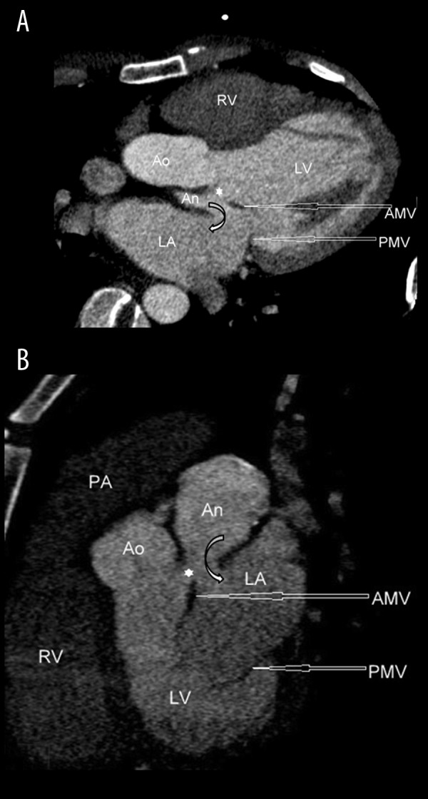
Figure 1.

(A, B) ECG-gated contrast-enhanced cardiac CT reformatted images showing a pseudoaneurysm [An] arising from the mitral aortic intervalvular fibrosa [*] and having fistulous communication with the left atrium [LA]. LV indicates the left ventricle. RV indicates the right ventricle. PA indicates the pulmonary artery. Ao indicates the aorta. AMV and PMV indicate the anterior and posterior leaflet of the mitral valve, respectively.
