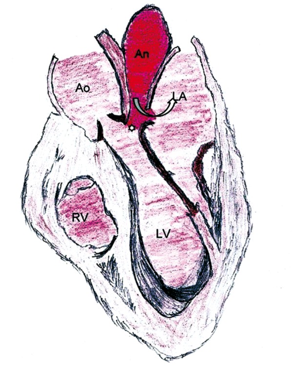
Figure 3.

A schematic diagram showing a pseudoaneurysm [An] arising from the mitral aortic intervalvular fibrosa [*], having fistulous communication [curved arrow] with the left atrium [LA]. Ao indicates the aorta. LV indicates the left ventricle. RV indicates the right ventricle.
