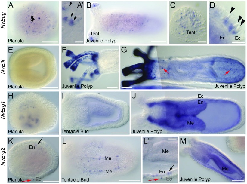
Fig. 2.
In situ hybridization analysis of Nematostella EAG family channel expression patterns. (A–D) NvEag expression is expressed in an ectodermal salt-and-pepper pattern in planula larva (A). Staining is adjacent to the cnidocyst capsules of the cnidocyte stinging cells (arrowheads, A′). NvEag expression is detected into juvenile polyps along the entire body axis, but is highly concentrated in the tentacle tips (B). Magnification of the tentacle tips reveals that NvEag staining in tentacles is also associated with cnidocyst capsules (C). Magnification of the polyp body wall shows that cnidocyst-associated expression is ectodermal (D, arrowheads). (E–G) NvElk expression is absent in planulae and during the early stages of polyp formation (E), but is elevated to high levels in polyp tentacles (F,G) and around the oral region (G). Some cells in the polyp body wall ectoderm also express NvElk (red arrows, G). (H–J) NvErg1 expression begins as punctae in the endoderm of planulae (H) and transitions to diffuse endodermal staining in polyps. (I,J) NvErg2 is expressed in a salt-and-pepper pattern in the ectoderm (red arrow) and endoderm (black arrow) of planula larva (K). Salt-and-pepper expression persists in endoderm (black arrow) and ectoderm (red arrow) through tentacle bud stage (L,L′). Expression appears slightly more concentrated around the mesentery structures (L). At juvenile polyp stages, punctate NvErg2 remains in the ectoderm, whereas endodermal expression becomes ubiquitous with individual cells enriched for NvErg2 (M). All images are lateral views with oral to the left and aboral to the right. Structures indicated are: tentacles (Tent.), endoderm (En), ectoderm (Ec) and mesenteries (Me). Scale bars: 100 μm (A,B,E–L,M); 10 μm (A′,C,D,L′).

