
Fig. 3.
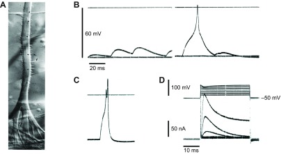
Electrogenesis in enzymically isolated smooth muscle from Beroe ovata. (A) Pharyngeal end of a living radial muscle cell. The fibre branches extensively; the endings anchor upon the epithelium, which is hidden by the pharyngeal circular muscles. Nomarski interference contrast. Scale bar, 20 μm. Note the multiple nuclei (arrowheads) and the thin sarcolemmal evaginations (arrows). From Hernandez-Nicaise et al. (Hernandez-Nicaise et al., 1980). (B,C) Series of spontaneous depolarizing events, possibly synaptic in origin, recorded from a fragment of longitudinal muscle produced by enzymic digestion (see Bilbaut et al., 1988a). In C, falling phase of the EPSP (and its short-circuiting effect) appears to end just before the peak of the action potential giving the appearance of a double spike. (D) Voltage-clamp records from same muscle fragment showing the typical response of a longitudinal muscle to depolarizing commands. Note that the clamp was imperfect at positive potentials because the micropipette could not supply the large currents necessary to control the membrane potential. Holding potential −50 mV; experiments conducted in artificial sea water at room temperature. Data from A. Bilbaut, M.-L. Hernandez-Nicaise and R.W.M., unpublished results.