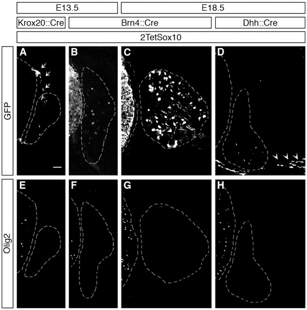
Fig 4. Oligodendrocyte-like cells in DRG do not arise from boundary cap cells, Schwann cells or peripheral neurons.
IHC was carried out with antibodies against GFP (A-D) or Olig2 (E-H) at E13.5 and E18.5 on transverse sections of embryos in which two copies of the TetSox10 transgene were activated by Krox20::Cre (A, E), Brn4::Cre (B, C, F, G), or Dhh::Cre (D, H) instead of Sox10::Cre. Contours of spinal cord and DRG are marked by stippled lines. Boundary cap cells are marked by arrows in A, Schwann cells by arrowheads in D. Size bar in A, valid for A-H: 50 μm.

