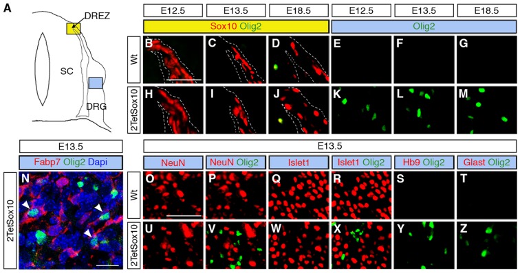
Fig 5. Ectopic Olig2-positive cells in the DRG express peripheral glial, but not neuronal or astrocytic marker and are not derived from boundary cap cells.
(A) Spinal cord (SC), DRG, dorsal root entry zone (DREZ) and peripheral nerves are schematically depicted. Areas shown in B-Y are marked as blue and yellow squares. (B-Z) Co-IHC was carried out on transverse sections (thoracic level) of wildtype (Wt, B-G and O-T) and 2TetSox10 (H-N and U-Z) embryos at E12.5, E13.5 and E18.5 with antibodies directed against Olig2 (B-N, P, R-T, V, X-Z, green), Sox10 (B-D, H-J, red), Fabp7 (N), NeuN (O, P, U, V), Islet1 (Q, R, W, X), Hb9 (S, Y) and Glast (T, Z). The areas from which pictures were taken are indicated by the blue or yellow color in the box above the panels. Color coding is according to the scheme in A. DREZ and spinal cord perimeter are demarcated by stippled lines in B-D and H-J. In N, nuclei were counter-stained with Dapi. Panels B-M and O-Z were photographed with a Leica DMI6000 B inverted microscope equipped with a Leica DFC350 FX camera, whereas panel N was taken with a Zeiss LSM 780 confocal microscope. Size bar: 50 μm in B and O (valid for B-M and O-Z, respectively); 20 μm in N.

