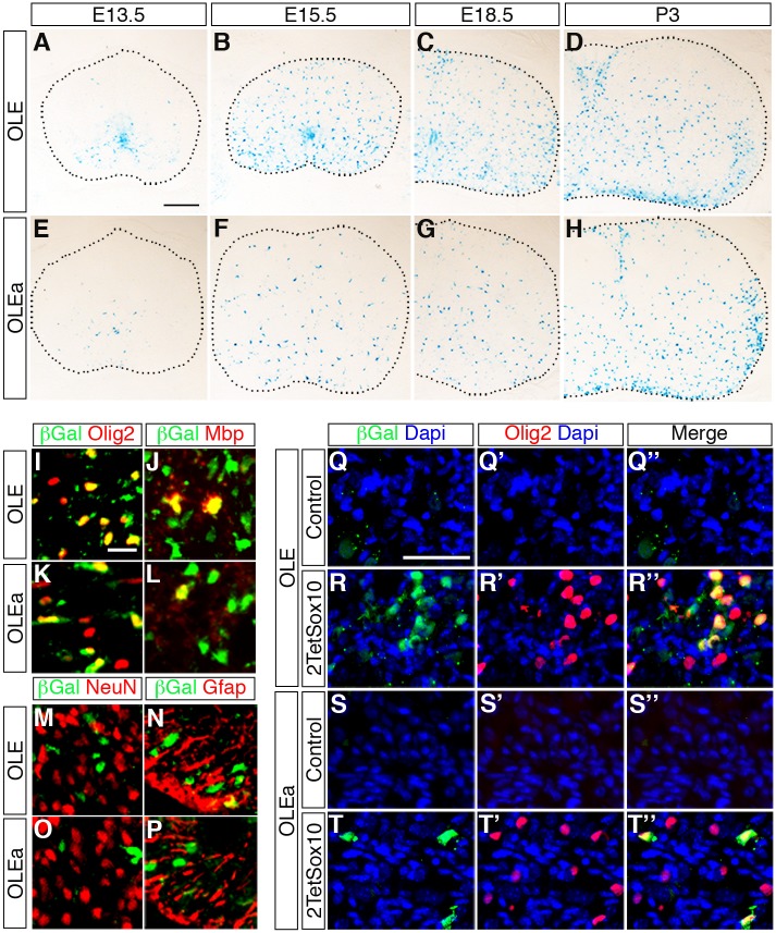
Fig 10. OLE is a Sox10-dependent oligodendroglial enhancer in vivo.
(A-H) The expression pattern of the lacZ reporter was followed during embryonic and early postnatal development in the spinal cord of OLE-lacZ (A-D) and OLEa-lacZ (E-H) transgenic animals by X-gal staining of transverse thoracic sections at E13.5, E15.5, E18.5 and P3. Size bar in A, valid for A-H: 200 μm. (I-P) Co-IHC was performed on spinal cord tissue of perinatal OLE-lacZ (I, J, M, N) and OLEa-lacZ (K, L, O, P) transgenic animals using antibodies directed against ß-galactosidase (in green) in combination with antibodies directed against Olig2 (I, K), Mbp (J, L), NeuN (M, O) and GFAP (N, P) (all in red). Pictures were taken from the ventral mantle zone for M, O and from the ventral marginal zone for all other panels. Size bar in I, valid for I-P: 20 μm. (Q-T”) Additionally, co-IHC was performed on DRG of mice that carried the OLE-lacZ (Q-R”) or the OLEa-lacZ (S-T”) transgene on a wildtype (Control) or 2TetSox10 background using antibodies directed against ß-galactosidase (green) in combination with antibodies directed against Olig2 (red). Nuclei were counterstained with Dapi (blue). Shown are single fluorescences for ß-galactosidase (Q-T) and Olig2 (Q’-T’) and the merge (Q”-T”) for each IHC. Size bar in Q, valid for Q-T”: 50 μm.

