
Fig 11. Olig2 overexpression does not generate oligodendrocyte-like cells in DRG.
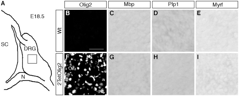
(A) Spinal cord (SC), DRG and peripheral nerves (N) are schematically shown at E18.5. (B-I) IHC and ISH were carried out on transverse sections (thoracic level) of wildtype (Wt) and 2TetOlig2 embryos at E18.5 with antibodies directed against Olig2 (B, F) and riboprobes against Mbp (C, G), Plp1 (D, H) and Myrf (E, I) and. Pictures were taken from the boxed DRG area. Size bars: 50 μm in B (valid for B-I).