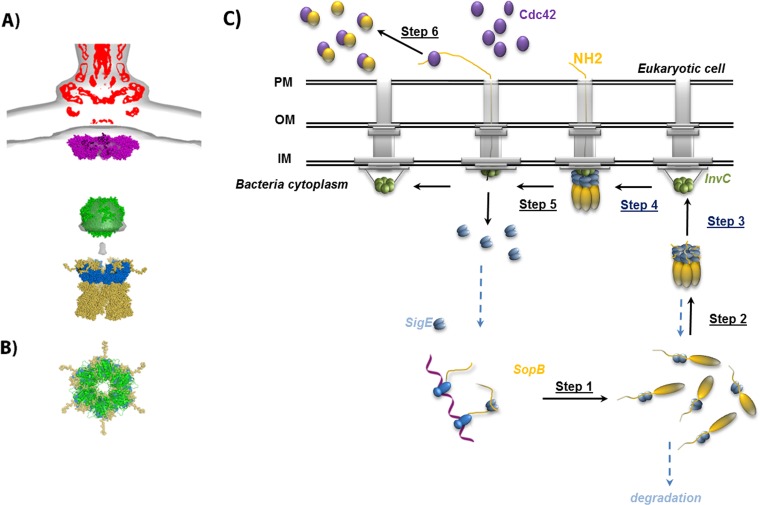
FIG 6.
Recognition and secretion mechanism of SopB. (A) Alignment of the model of SigE/SopBΔ29 (blue and yellow) with the superposition of the cryo-electron microscopy density map of isolated (EMD-1875; red) and in situ (EMD-2521; gray) needle complexes in which models of MxiA, a homolog of InvA (magenta; PDB ID 4A5P), and the hexameric ring model of FliI ATPase, a homolog of InvC (green) are included, as described in reference 8. (B) Superimposition of the SigE/SopBΔ29 and ATPase models along the axis of the central tunnel of their ring with the same color code as in panel A. (C) The proposed mechanism explains the injectisome recognition by the SigE/SopB complex and the translocation initiation of this complex through the T3SS (see the text for more details).

