
FIG 7.
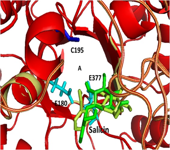
The overlaid structures of wild-type (yellow) and mutant (red) BglA proteins. The structure of the wild-type protein is based on PDB ID 2XHY (8), and the structure of mutant BglA was modeled using the Rosetta software with 2XHY as the template. The letter A indicates the approximate substrate binding pocket. The position of C195 in the wild-type structure is highlighted in blue, and the active-site glutamate residues E180 and E377 are shown in cyan. Salicin docked in the binding pocket is shown in green for the wild-type structure and in lemon for the mutant.