
Figure 2.
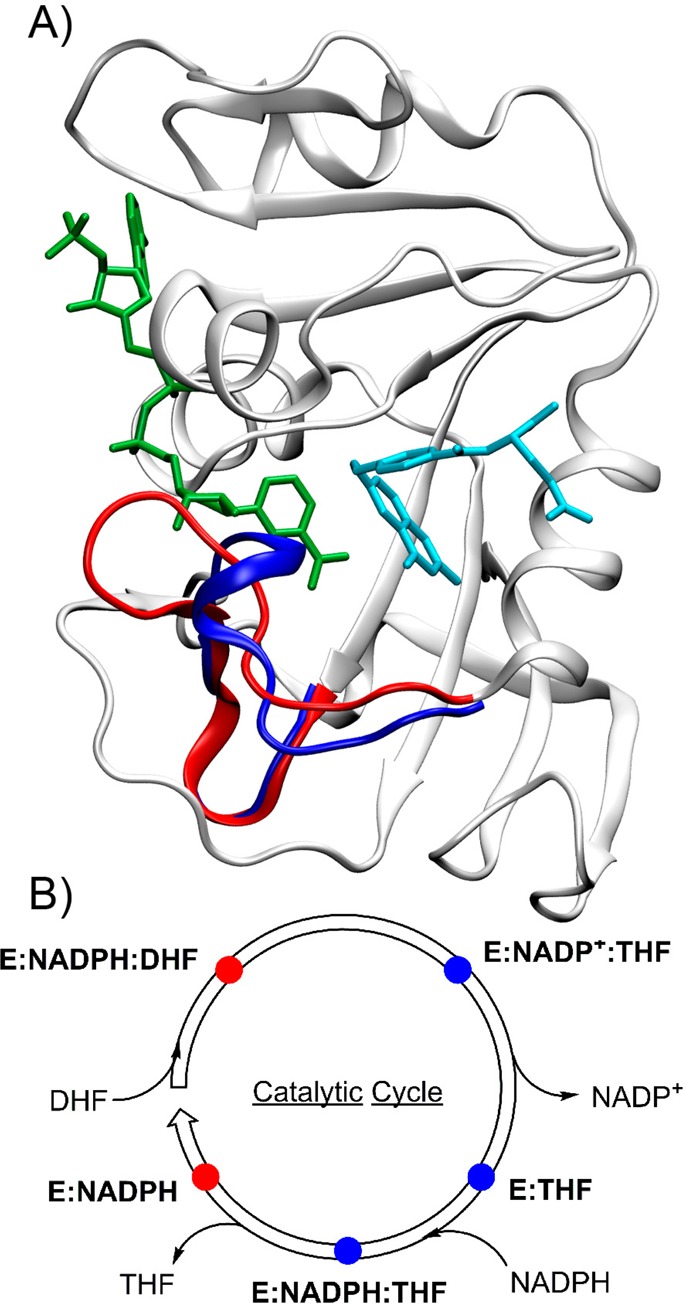
(A) Ribbon structure of ecDHFR with bound NADP+ (green) and folate (sky blue) illustrating the differences between the closed (PDB ID 3QL3; red) and occluded (PDB ID 1RX6; blue) conformations of the Met20 loop. (B) The ecDHFR catalytic cycle distinguishing the closed conformation (red) and occluded conformation (blue). Adapted with permission from ref (10). Copyright 2014 American Chemical Society.