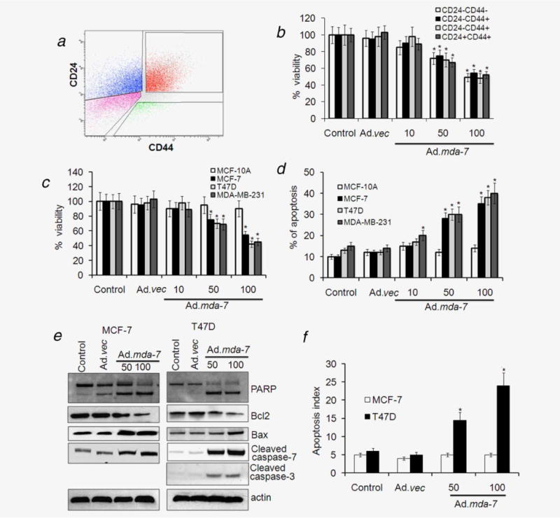
Figure 2.

Ad.mda-7 infection induces apoptosis in breast cancer-initiating/stem cells. (a) MCF-7 cells were analyzed by flow cytometry for CD44 and CD24 expression. The gates shown were used for sorting of cells with the indicated phenotypes. (b) The sorted populations, based on surface markers, were infected with Ad.mda-7 (10, 50 and 100 pfu/cell) or Ad.vec (100 pfu/cell) and after 48 hr cell proliferation was measured by MTT assay. (c and d) Cancer-initiating/stem cells from MCF-7, T47D, MDA-MB-231 and MCF-10A were infected with Ad.mda-7 (50 or 100 pfu/cell) or Ad.vec (100 pfu/cell) and after 48 hr cell proliferation was measured by MTT assay (c) and Annexin V staining assay using flow cytometry (d) measured apoptosis. MCF-7- and T47D-initiating cells were infected with Ad.mda-7 (50 or 100 pfu/cell) or Ad.vec (100 pfu/cell) and cell lysates were prepared after 48 hr. (e) MCF-7- and T47D-initiating cells were infected with Ad.mda-7 (50 or 100 pfu/cell) or Ad.vec (100 pfu/cell) and cell lysates were prepared after 48 hr. Activation of poly(ADP-ribose) polymerase-1 (PARP), caspases 3 and 7 and expression of Bcl-2 and Bax were detected by Western blotting analysis. (f) After 48-hr infection with Ad.mda-7 or Ad.vec the apoptotic index as indicative of caspase 3/7 activity in MCF-7- and T47D-initiating cells was determined using a fluorescence microscope. The apoptotic index was calculated by dividing the number of caspase 3/7 fluorescent cells by the total number of DNA-containing cells after staining with Vybrant DyeCycle Green. Data represent mean ± SD (n = 3). Asterisk (*) represents significant difference (p < 0.5) with corresponding control.
