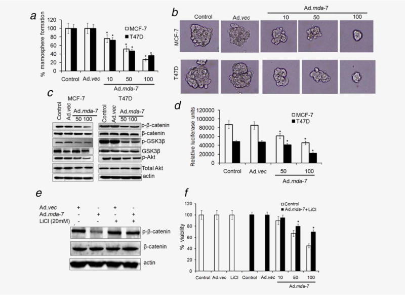
Figure 4.

Ad.mda-7 inhibits mammosphere formation and Wnt signaling in breast cancer-initiating/stem cells. (a) Cancer-initiating/stem cells from MCF-7 and T47D were infected with Ad.mda-7 (50 or 100 pfu/cell) or Ad.vec (100 pfu/cell) for 48 hr and the infected cells were seeded for formation of secondary mammospheres as described in “Material and Methods” section and quantified by counting the mammospheres by microscope. Data represent mean ± SD (n = 3). (b) Photomicrograph of primary mammospheres following the indicated treatments (magnification, ×100). (c) MCF-7- and T47D-initiating/stem cells were infected with Ad.mda-7 (50 or 100 pfu/cell) or Ad.vec (100 pfu/cell) and cell lysates were prepared after 48 hr and changes in phospho- and total β-catenin, GSK3β and Akt expression were evaluated by Western blotting. (d) MCF-7- and T47D-initiating/stem cells were infected with a TCF/LEF TOP-luc lentiviral reporter system for 48 hr followed by infection with Ad.mda-7 (50 or 100 pfu/cell) or Ad.vec (100 pfu) for 12 hr and relative luciferase activity was measured as described in the “Material and Methods” section. (e and f) T47D-initiating/stem cells were infected with 100 pfu/cell of Ad.mda-7 or Ad.vec in the presence of LiCl (20 mM) for 48 hr and expression of phospho- and total β-catenin was analyzed (e) and cell proliferation was measured by MTT assay (f). Asterisk (*) represents significant difference (p < 0.5) when compared to Ad.mda-7-infected cells without LiCl.
