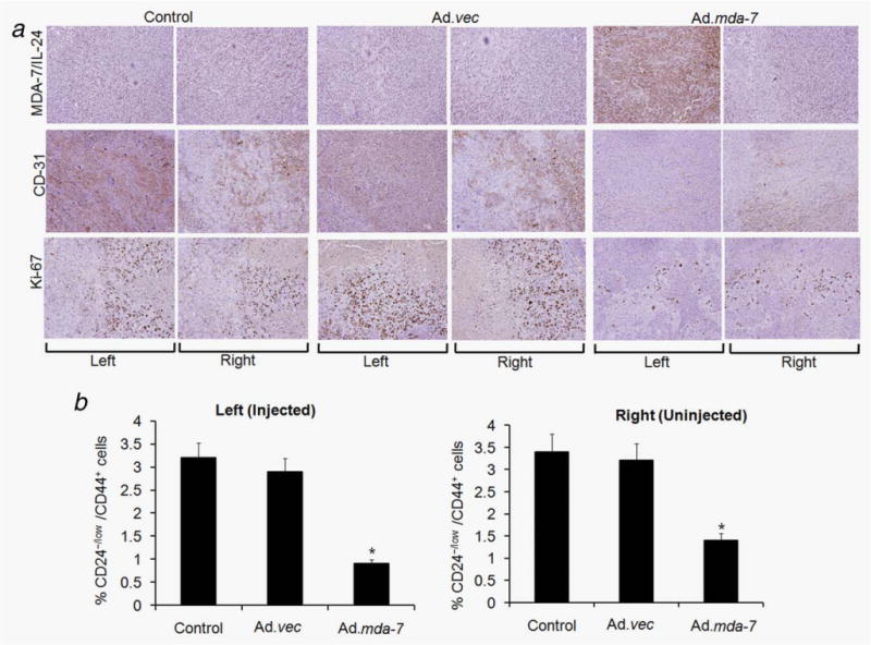
Figure 6.

Immunohistochemical analysis of tumor xenografts induced by T47D-initiating/stem cells infected with Ad.vec or Ad.mda-7 or controls injected with PBS. Tumor tissues were harvested and formalin-fixed and paraffin-embedded sections were immunostained for MDA-7/IL-24, CD-31 and Ki-67. The left and right tumors were from animals injected three times (left tumor) with PBS, Ad.vec or Ad.mda-7. Experimental details are given in “Material and Methods” section. (b) Single-cell suspensions were generated from the tumors of all groups and were fixed and stained with CD24 and CD44 antibodies to quantify the percentage of CD24−/low/CD44+ cells by fluorescence-activated cell sorting. The data represent the mean ± SD with a minimum of five mice per group. Asterisk (*) represents significant difference (p < 0.5) vs. corresponding control.
