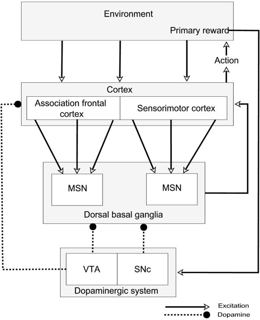
Figure 1.
Some key anatomical components of the corticostriatal loops involving cognitive and motor areas of the dorsal striatum. Dopaminergic neurons of the SNc are lost in Parkinson’s disease, with relative sparing of the dopaminergic neurons of the VTA. In the model, the dorsal basal ganglia is represented by direct pathway medium spiny neurons. MSN – striatal medium spiny neuron; SNc – substantia nigra pars compacta; VTA – ventral tegmental area.

