
Figure 4.
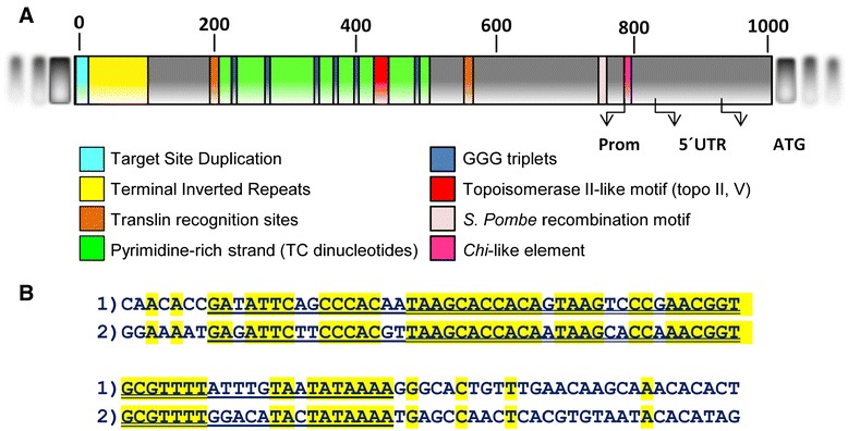
Conserved motifs in the Cit_Mule_1 terminal region. A) Schematic representation of the terminal 5′ sequence of Cit-Mule_1 showing the TSD (light blue), a fragment with the terminal inverted repeats (yellow), two translin recognition sites (brown), TC stretches (green), GGG triplets (dark blue), a putative topoisomerase II-like motif (red) and two specific DNA motifs of recombination hot spots identified in Schizosaccharomyces pombe (pink) and Escherichia coli (magenta). Arrows show the first nucleotide of the promoter, the first one of the 5′UTR and the first ATG of the protein according to the ORF prediction. B) Sequences of the 100 terminal-most nucleotides of CitMule_1. The “1” sequence represents the upstream terminal end of CitMule_1 and the “2” sequence the downstream end. The double-underlined sequences indicate tracks with 88% similarity, while the underlined sequences denote tracks with lower similarity (78%).