Fig. 1.
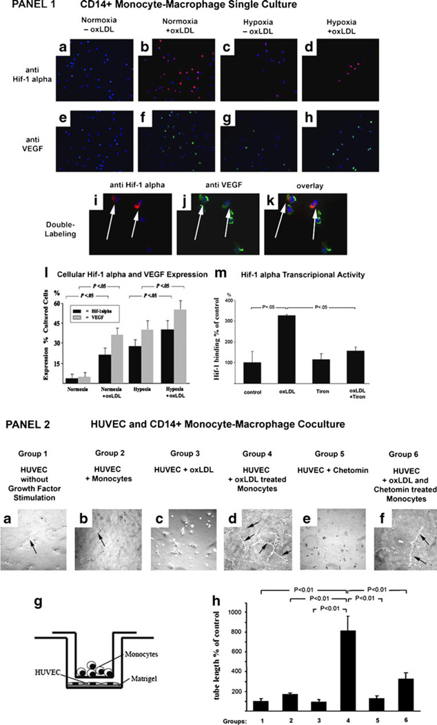
Panel 1: Effect of oxLDL on HIF-1α and VEGF in monocytes. a–h Representative pictures of HIF-1α (a–d) and VEGF (e–h) expression in monocytes cultured under normoxia without oxLDL (a, e), under normoxia with oxLDL (b, f), under hypoxia without oxLDL (c, g), and under hypoxia with oxLDL (d, h). A strong induction of HIF-1α and VEGF by oxLDL was observed independent of the presence of hypoxia (×400). i–k Double labeling for HIF-1α (i) and VEGF (j) showed clear co-localization of both antigens in single oxLDL-treated monocytes (overlay in k) (× 1,000). l Quantitative evaluation confirmed the significant increase in HIF-1α and VEGF expression with oxLDL treatment to a degree similar to hypoxia-induced expression. Combining oxLDL and hypoxia further enhanced the expression of both HIF-1α and VEGF (ANOVA, P<0.05). m Transcriptional activation of HIF-1α as measured by the Trans-AM assay was elevated significantly in monocytes treated with oxLDL. Of note, co-treatment with the antioxidant (tiron) abrogated HIF-1α transcriptional activation induced by oxLDL (ANOVA, P<0.05). Panel 2: Effect of oxLDL on monocyte-mediated angiogenesis. a–f HUVECs were seeded onto growth factor-depleted Matrigel in a transwell co-culture setup (shown in g) in the presence of monocytes (b, d, f) or without monocytes (a, c, e). The monocytes were either left untreated (a, b), treated with oxLDL (c, d), treated with the HIF-1α inhibitor chetomin (e), or with oxLDL and chetomin (f). HUVECs grown alone on growth factor-depleted Matrigel did not show significant tube formation (a) and were used as negative control. HUVECs under the same conditions grown in the presence of untreated monocytes did not show tube formation either (b). Likewise, HUVECs grown alone but treated with oxLDL did also not show signs of tube formation (c). However, HUVECs grown in the presence of oxLDL-treated monocytes did show extensive tube formation (arrows) reflecting increased angiogenic activity (d). Of note, co-treatment of monocytes with the HIF-1α inhibitor chetomin significantly suppressed the proangiogenic effect of oxLDL treatment (f) and chetomin did not have an effect on HUVECs alone (e). g Schematic drawing of the monocyte endothelial co-culture system for determining capillary tube-like structure formation h Bar graphs comparing the length of endothelial tube formation as measured in micrometers. In summary, groups 1–3 and 5 served as various controls, and groups 4 and 6 were the two treatment groups as described from a to f (ANOVA P<0.01; error bars ± SEM)

