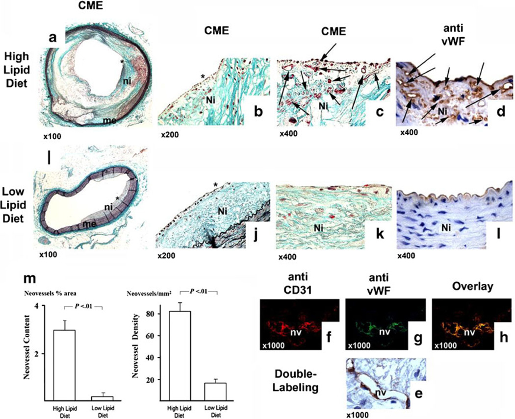
Fig. 3.
Effect of dietary lipid lowering on in vivo angiogenesis in experimental atherosclerosis, a–l Representative pictures of the effect of dietary lipid lowering on intimal neovascularization in a rabbit atherosclerosis model (a–d arteries of animals with high-lipid diet; i–l arteries of animals on low-lipid diet). With increasing magnification, the extent of neovascularization (arrows) especially in shoulder areas (asterisk) of large and foam cell-rich atherosclerotic lesions of animals with hyperlipidemia becomes visible on serial sections stained with Masson Trichrome and vWF antibody (a–d). In contrast, after normalization of serum lipids, the microvascular structures are nearly completely absent from a largely fibrotic intima and vWF immunostaining is only seen at the luminal endothelial layer (i–1). m Bar graphs comparing neovessel content (percent intimal area covered with neovessels) and neovessel density (intimal neovessels per square millimeter) with high- vs. low-lipid diet. Hyperlipidemia resulted in strikingly enhanced neovascularization compared to animals with normolipidemic conditions (independent sample t test, P<0.01; error bars ± SEM). f–g High power magnification showing double immunofluorescence labeling for CD31 (f) and for vWF (g) with significant overlay (h) of both signals confirming the endothelial lining of microvascular structures of hyperlipidemic animals.

