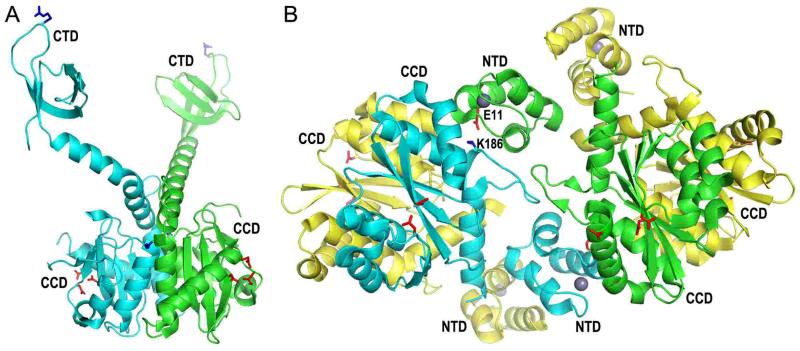
Figure 3.
Structures of 2-domain HIV-1 integrase constructs. (A) The X-ray crystal structure of the HIV-1 integrase CCD-CTD dimer (pdb code 1EX4) (66), highlighting the CCD and CTD side-chains that were shown in Fig. 2. (B) The crystal structure of the NTD-CCD asymmetric unit (pdb code 1K6Y) (149) highlights NTD residue Glu11 and CCD residue Lys186 of the green and cyan molecules, respectively, which play important roles in integrase concerted integration activity and HIV-1 infection (70). The other pair of interacting residues (Glu11 from the cyan NTD and Lys186 from the green CCD) is not visible in this projection. The side chains of the DDE catalytic triad (red sticks) and NTD-coordinated zinc atoms (grey spheres) are also shown.

