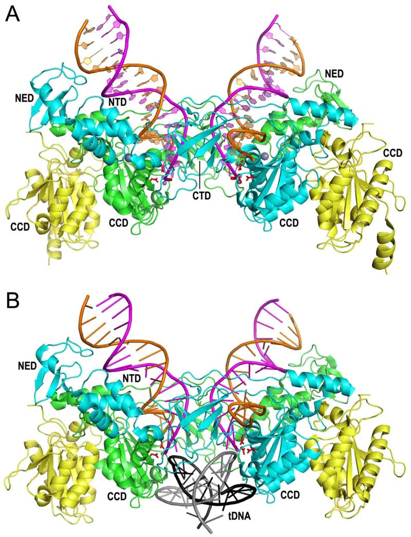
Figure 5.
PFV intasome structures. (A) Structure containing 19 bp pre-cleaved U5 DNA end (94, 207) (pdb code 3OY9). The inner integrase monomers of the tetramer, which contact the viral DNA, are painted cyan and green; the outer integrase molecules are yellow. The transferred DNA strands with CAOH 3′ ends are painted magenta whereas the non-transferred strands are orange. The large grey spheres are NTD-coordinated zinc; small grey spheres are Mn atoms coordinated by the DDE active site residues (red sticks) and viral DNA end. (B) Structure of the TCC (98) (pdb code 3OS1). Although a 30 bp target DNA (tDNA) was utilized during crystallization, only 18 bp (grey plus strand sequence -7GCACGTG\CTAGCACGTGC10) was resolved in the electron density maps.

