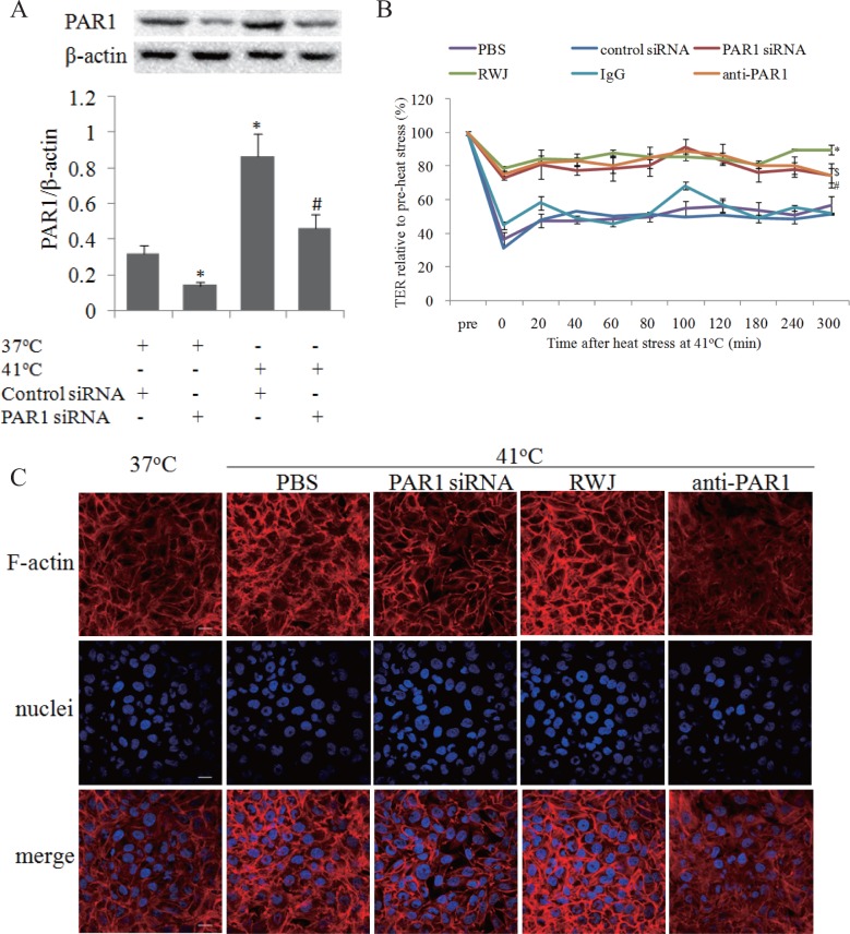
Fig 3. PAR1 mediated endothelial hyper-permeability and F-actin rearrangement induced by heat stress.
Mono-layer HUVECs were treated with RWJ56110 (RWJ; 5μM), PAR1 neutralizing antibody (anti-PAR1), control IgG (IgG), PAR1 siRNA, or negative control siRNA, followed by heat stress at 41°C for 2h. Mono-layer HUVECs were transfected with control siRNA or PAR1 siRNA respectively, or treated with RWJ, anti-PAR1 and IgG, followed by heat stress at 41°C or cultured at 37°C for 2h, PAR1 protein expressions were determined by western blot. (a) Representative images of western blot and quantitative analysis of PAR1 protein normalized to β-actin were shown (n = 4, * vs. PBS group, P < 0.05). (b) TER was determined similar to Fig. 1A (n = 4, *P < 0.05, vs. PBS group, $ vs. control siRNA group, # vs. IgG group, P < 0.05). (c) Following above heat stress, the cells were recovered at 37°C for 2h, and nuclei (blue) and F-actin (red) were stained as Fig. 1B noted, scale bar: 20μm.

