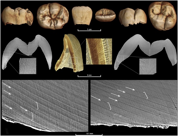
Fig 1. Multiscale synchrotron imaging of a fossil Homo juvenile individual.
DNH 67 (right lower first molar: left), DNH 71 (right upper central incisor: middle), and DNH 70 (left upper first molar: right). Images are from scans performed with the following voxel sizes: 20 μm (upper row), 5 μm (middle row), and 0.7 μm (lower row; DNH 67: left, DNH 70: right). An identical internal developmental defect pattern confirmed that the two molars, which were found isolated but in close proximity, came from the same individual. They also both show an identical long-period line periodicity of 8 days, as 8 light and dark bands (cross-striations illustrated in white brackets) can be seen between successive long-period lines (Retzius lines illustrated by white arrows). It was not possible to determine the age death for this individual due to postmortem loss of the incisor cervix and dentine from all teeth.

